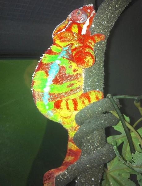
WARNING, GRAPHIC PHOTOS—Necropsy of Panther with severe nutritional MBD

Dr O

Veterinarian
A friend that owns an exotics pet shop sent me this photo and video of a Panther Chameleon donated to him by an owner who claimed that "he was doing everything right" but the lizard was acting sick. After seeing the video and the severe distress that the cham was in, I told my friend that I would take it and try to nurse it as best as I could, but the prognosis was exceedingly grim from just the video evidence that I was presented with. Upon receiving him, the physical exam revealed several long bone and joint fractures that were fresh or unhealed, severe edema around his neck, distal limbs, ventral chest and abdomen, curved and soft long bone formation, severe bilateral exophthalmos (eyes pushed outwards from the socket) with ocular discharge, grasping at his own limbs, and periods of tetanic seizures and gaping behavior. However, the most worrisome sign was a very gritty feel upon palpation of his liver, mesentery, and intestinal tract. I performed a quick fine needle aspirate of his peritoneum which should basically be an empty vacuum, but indeed received a blood-tinged and flocculent (suspended particles) fluid in the syringe. Typically I would immediately fix, stain and dry the aspirate to look under a microscope at the cells that I had, but I was unfortunately out of the necessary stains. However, I knew at that point that his prognosis was terminal. At the time another forum member just happened to be over and asked an excellent question, did I think that the gritty feeling was possibly from calcification of the soft tissues from oversupplementation of calcium? However I said that it was unlikely as the gritty texture appeared to be on the serosal (external) surfaces of the organs and was more likely from inflammatory cells such as fibrin and blood clots from the fluid in the abdomen. Organ calcification tends to distribute evenly throughout the tissue and not just on the outside of the organs. With that, I instituted therapy with Repashy RescueCal+, high dose enrofloxacin, tobramycin ophthalmic ointment, oral fluids supplemented with Reptisafe and Reptaid, full day sunlight exposure in a screened cage, and elevated temperatures during the nights. With all of my hospital equipment still in storage several states away, I was not able to place a catheter as I would have normally tried to do for direct intraosseous fluids and injectable medications (however, the necropsy revealed that placement of a catheter would have been difficult to impossible due to the destuction of most long bones). By the fourth day he was worsening, only able to lay on the floor of his cage or hang upside down from his rear feet, but he had obviously become much weaker. As I felt that he had a terminal situation which was worsening and that he was suffering, I chose to euthanize him with Beauthanasia-D. Immediately afterwards I performed a necropsy and photographed the procedure for the members of this forum. In the photographs below you will see some examples of how nutritional MBD can wreak havoc upon the body, as well as normal anatomical structures which some may find interesting.


First, the photo and video that was emailed to me by the pet shop owner. You can see the bulbous eyes with drainage, some curvature to the limbs, self-grasping behavior and involuntary muscle movements.:

full


http://youtu.be/EZphRnqEJx0


After the euthanasia, his body was laid out on a canine Wee-Wee pad which was excellent at absorbing fluids without leting them pass through the material.

full


full


full


His left forelimb was dissected down to the bone where a broken and displaced radius is visible (A) in addition to a metacarpal (wrist) joint that was mobile in all directions from the complete destruction of the joint (B). Notice how it is easily reflected backwards in the second photo.

full


full


The right forelimb had an unhealed fracture of the ulna which almost gave the appearance of him having "two elbows". (A) is where the elbow joint is, and (B) is where the fracture exists. Note in the second photo how there is flexion in both regions. The brachial vein (red) and artery (white) are clearly visible in the upper part of the limb as well.

full


full


Removal of the muscle and connective tissue shows a short, stunted and curved radius with the thickened fracture of the ulna. Throughout the necropsy, the bones were soft and rubbery as a chicken bone becomes after soaking in vinegar for several days.

full


The oral cavity was surpsingly clean. The first photo shows the normal position of the tongue as it leaves the hyoid bone (A). Note the various muscles and blood vessels within the tongue

full


A typical visualization of the open glottis (A) which leads to the trachea (B) and then to the lungs. This would be where an endotracheal tube would be placed to allow oxygen and anesthetic gasses to be delivered to the lungs. As its normal position is at the bottom of the oral cavity and it has a strong flap of muscles that can close it, one can see how it is quite easy to tube feed or give oral medications to most reptiles without fear of getting any in the windpipe.

full


Both hemipenes (A) were everted and appeared normal other than the large waxy plugs (B) that were inside each hemipene.

full


(To be continued; please do not post until entire article is completed)
 
Last edited by a moderator:
The next set of photos shows the skin reflected off of the ribcage before the coelomic cavity is revealed. Once it is, the advanced degree of coelomitis is seen. Organs visible at this point include (A) the heart inside of the opened pericardial sac, (B) the liver with a cobbled surface, (C) the small intestines which appear blackened and also have inflammatory cells on the serosal surfaces, (D) the colon which appears normal, (E) a testicle, and (F) the aorta which can be followed in several of the photos. However, the red-tinged fluid throughout the cavity is completely abnormal and is what was aspirated while he was still alive.


full


full


full


After the fluid was blotted up as much as possible, the organs can be seen more clearly. Here we have (A) the exposed heart, (B) a lung, and (C) the aorta. Between the heart and the liver many strands of fibrin and scar tissue exist.

full


A normal appearing chameleon heart with its one ventricle (A) and two atria (B).

full


The lungs (A) of chameleons are exceedingly simple in organization and structure, and this lung was on the verge of complete destruction from the exposure to the fluid that was surrounding it in addition to the likely toxins and bacteria that were building up in his system. Just handling it gently with surgical instruments caused it to shred and fall apart.

full


The mid and caudal abdomen when cleaned show (A) the esophagus and how it widens into (B) the stomach. (C) is a normal colon (large intestine). (E) is a testicle and (D) is a portion of the left kidney. This is one of the best views of the aorta and if you look closely, many branches into the intervertebral spaces and other organs can be made out.

full


The complete gastrointestinal system from (A) the esophagus, (B) the stomach, (C) the small intestine, and (D) the colon. The SI in chams is often blackish in color but this appears to be excessively so. Possibilities for the color change inculde cellular death of the SI, bleeding into the lumen of the SI, and color change from the oral medications. Likely it is a combination of all three factors.

full


The next three photos show most of the urogenital system in addition to (A) the spleen. (B) are the testicles, (C) are the kidneys, and (D) are the vas deferens which carry the sperm from the testicles to the hemipenes. The photos are in sequence from one side of the body, and the organs are flipped over to show the bilateral view, and then finally the opposite side of the body. (E) the aorta and (F) the vertebra are seen in the final photo.

full


full


full


An incised and opened testicle showing the sperm contained inside (A).

full


A head-on view of the sectioned kidney. The kidneys were grossly swollen with destruction of the normal architecture, so he was in kidney failure along with everything else.

full


The heart with the ventricle (A) and atria (B) transected. There were no abnormalities to be seen with the naked eye.

full


(to be continued; please do not post until the article is finished)
 
Last edited by a moderator:
At this point the main part of the necropsy was finsished with several diseased organs noted on gross exam, and histopathology of the organs would have seen a tremendous amount of microscopic damage. I continued on just for the sake of anatomy. Many people have never seen a chameleon eyeball with the eyelids (turret) removed, and the globes can be seen in the following shots on both sides of the body. However, whereas the eyeballs themselves appeared normal, the orbital space where the globes live were much too shallow and can be best seen in the second photo. This explained the exophthalmos (protruding eyes). Essentially his bones were so deformed that even his skull, zygomatic and orbital bones were affected to the point where his eyeballs could not fit inside of the skull anymore.


full


full


full


full


full


Finally I decided to expose the brain, as many people wonder just how big it is. I had to strip all of the tissue and muscles from the skull and began by entering a caudal portion of the skull. After delicately manuvering around, I was able to find one of the large optic nerves (A). At that point I could trace it backwards as the optic nerve, also known as cranial nerve II connects to the forefront of the brain. Dissecting back further brought me to the optic chiasm which is where the nerves cross from one side of the body to the other at this junction ("the right eye is seen by the left brain, and the left eye by the right brain"). It is also interesting to note that the nerves are quite large for the size of the animal; demonstrating the importance of vision in a chameleons life.


full


full


full


And finally the brain can be seen (B) just behind the optic chiasm (A). Not much there, just the basics. Food, shelter and sex make up the reptilian brain, and there you have it.

full


On reflection of the case I would not have done anything differently. From his initial condition, examination and few diagnostic tests I was able to determine that what he had was a terminal prognosis. And even if by some miracle he would have been able to pull through, he was living a life where his bones were so damaged that his eyes could not even fit in their sockets anymore, nevermind the kidney failure, multiple fractures, severe infection, early pneumonia et al. There would not have been any quality of life in my opinion and I am glad that he was able to pass easily. As most of us know who own chameleons, they need light X, diet Y, and temperature/humidity Z. If they do not have at least these three conditions met to an exacting degree, this may be the end result. I hope that you have learned something that you did not know before, and of course I hope it helps prevent future cases of an entirely preventable situation.


dr. o—
 
Last edited by a moderator:
It was difficult for me to examine the entire necropsy. But it was very informational. Thank you for posting this and thank you for ending the poor chams life.

I hope this will make more people aware of what we do to our chams if they do not get proper care.
 
im not sure if its just me but im unable to see any pictures only the video at the begining
 
I was wondering the same thing - anatomy has always fascinated me . . .

That poor guy looked young - I guess he could be just small from not being well taken care of - but he is about the size of my 7 month old . . .
 
very informative. alot of people cant stomach to do something like that but i'm glad there are those out there who can because having a better understanding of things like this makes life for other chameleons out there just a little bit better
 
one other finding, or lack of, to mention. chameleons tend to store very firm, heavy and discrete fat bodies in their caudal abdomen. a typical size fat body in a cham his size would be 1"x2", and there should be several. needless to say not a gram of fat was noted during the necropsy. that does not necessarily mean that he was not fed, rather he was completely unable to convert any nutrition into fat storage since virtually everything in his body had gone haywire. there was even a (almost undigested) cricket in his colon, and in the video they mention that he was apparently still eating.
 
You mention about the idea of over supplementing with calcium leading to calcification of the soft tissues. This is something I haven't yet read about on the forum but something that is interesting to think about after seeing a number of calcifications within human radiographs. Is this a large concern when it comes to supplementing with calcium? I've read a number of forums about supplementation including one of yours so I am doing my best to become as familiar with a routine as possible, but I am still very new! (In fact, my female veiled chameleon named Aloe has started to shed for the very first time since I got her a month ago today! :))
 
You mention about the idea of over supplementing with calcium leading to calcification of the soft tissues. This is something I haven't yet read about on the forum but something that is interesting to think about after seeing a number of calcifications within human radiographs. Is this a large concern when it comes to supplementing with calcium? I've read a number of forums about supplementation including one of yours so I am doing my best to become as familiar with a routine as possible, but I am still very new! (In fact, my female veiled chameleon named Aloe has started to shed for the very first time since I got her a month ago today! :))

absolutely, abnormal overuse of calcium/vit. D/and UVB can lead to calcification of soft tissues which is often irreversible. i want to be clear, this is a possibility when mega-doses are being used in treatment of a calcium disorder. the kidneys tend to be the initial organs that are damaged, but it can lead anywhere. primary renal disease, hyperparathyroidism, and diets too high in protein can also be causes. additionally, calcification of soft tissues is only one of the problems with hypercalcemia; other concurrent problems tend be even more serious in their nature.
 
Last edited:
Dr. O, thank you for doing this. I'm a wimp so I didn't want to see the pictures, but I was very interested in reading your comments. If there are other wimps, right click on the first post in the thread, select "view source"....scroll past all the pretty colored stuff and read the blocks of black typing.

in your opinion, is there, in fact, a danger of over supplementing with calcium?

**edited**

Oops...you posted while I was composing.
 
You mention about the idea of over supplementing with calcium leading to calcification of the soft tissues. This is something I haven't yet read about on the forum but something that is interesting to think about after seeing a number of calcifications within human radiographs. Is this a large concern when it comes to supplementing with calcium?

additionally, all of the aforementioned diseases can cause this in humans as well, but many calcifications on radiographs may also just be the result of some scar tissue formation within the organ. for instance, after an extensive backpacking trip through SE Asia for 6 months I came home with a visible calcified lesion in my lower left lung. likely the result of a walled-off infection or parasite. but it has remained the exact same size for over 10 years and neither my doctors or myself are concerned about it. something like that may be commonly seen on radiographs of any creature. but if it's a disease process, the lesions will grow and/or multiply. and continued oversupplementation of minerals and vitamins would do that as well.
 
Back
Top Bottom